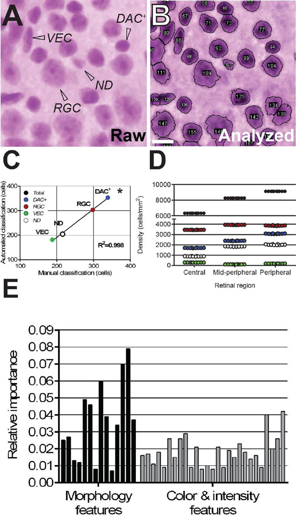
Figure 1. RetFM-Class performs automated cell type classification with an accuracy and precision similar to those of manual classification.
(A) Representative image from an H&E-stained retinal whole-mount containing three categories of distinguishable cells (RGC, retinal ganglion cells; VEC, vascular endothelial cells; DAC+, displaced amacrine cells plus other cells with similar-appearing nuclei), as well as a fraction of what appear to be nuclei but are difficult to assign to another cellular category manually because their features are not distinct (ND). (B) Automated nucleus recognition and counting of the same image shown in panel A by RetFM-J. (C) Comparison of manual- and auto-classification (n=10 images; 1,041 nuclei). (D) Comparison of 10 repeat classifications of cells with RetFM-Class from an individual image. Cell type is color coded as in panel C for RGC (red), DAC+(blue) VEC (black), and ND (open). Note that the Y-axis is discontinuous and not equally scaled in the upper and lower portions. (E) Auto-classifications by RetFM-Class were multifactorial, using multiple image features related to morphology and color and intensity to varying extents. In consideration of space, labels for individual bars may be found in Supplemental Table 1, with the order of the features in the bar graph (left to right) matching the presentation in the Table (top to bottom). Scale bar = 10 µm.

