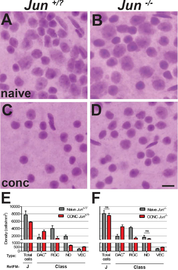
Figure 4. RetFM-J, but not RetFM-Class, performance matches expectations in acute damage induced by CONC.
Mice of the same cross and genetic background. Jun+/? mice (left column) undergo apoptosis in response to CONC. Jun−/− mice (right column) have resistance to CONC-induced apoptosis but are capable of pre-apoptotic changes such as nuclear atrophy. In the naïve control groups, retinas from (A) Jun+/? and (B) Jun−/− retinas both exhibit dense populations of cells of varying type in the inner retina. In the CONC-injured groups assessed 35 days after induction, (C) Jun+/? retinas have decreased cell density and the majority of remaining cells have small, densely stained nuclei. (D) Jun−/− retinas have nearly normal cellular density and the majority of remaining cells again appear to have small, densely stained nuclei. Note that the diversity in appearance of nuclei is greater in panel D than C. Scale bar = 10µm. – Bar graph of cell density versus cell category. RetFM-J detected a loss of total cells in Jun+/− and not Jun−/− retinas, matching expectations, but RetFM-Class predicted statistically significant differences between CONC and naïve mice in nearly all cellular categories of both genotypes, indicating that classification performance was negatively influenced by the induced synchronous insult of CONC (p<6.1E−3, Bonferroni-corrected 2-tailed Student’s t-test for all comparisons except those noted with ns). Note that the Y-axis is discontinuous and not equally scaled in the upper and lower portions. Jun+/? naïve (n = 8 retinas, 269,743 nuclei), CONC (n = 4 retinas, 75,589 nuclei); Jun−/− naïve (n=6 retinas, 157,735 nuclei), CONC (n=4 retinas, 100,410 nuclei); mean ± 1 SD.

