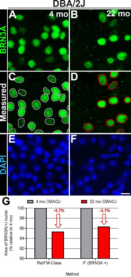
Figure 7. Confirmation that surviving retinal ganglion cells in DBA/2J mice with glaucoma have smaller nuclei.
Immunofluorescent images from the inner retina of whole-mount preparations showing labeling of BRN3A–positive nuclei of presumptive retinal ganglion cells in (A) pre-diseased DBA/2J mice at 4 months of age (left column) and (B) glaucomatous DBA/2J mice at 22 months of age (right column). Note that there are decreased numbers of BRN3A–positive nuclei in retinas from the older DBA/2J mice with glaucoma with age and onset of glaucoma. The same fields are also shown below with (C, D) nuclear outlines indicating the areas that were measured and with (E, F) DAPI-labeling. (G) Graph comparing the change in mean area of DBA/2J nuclei (as a percentage relative to that at 4 months) in glaucoma as measured by RetFM-class (left; n = 99,629 nuclei at 4 months of age; 27,732 nuclei at 16 months of age) and immunofluorescence (right; n = 2,369 nuclei at 4 months of age; 1,449 nuclei at 22 months of age). Note that both methods reveal a similar 3.7–4.7% reduction in the mean nuclear area of retinal ganglion cells. Scale bar = 10 µm.

