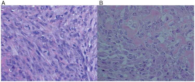
Figure 1.

(A) Photomicrograph of tumour histology from mouse receiving UWKOS3. The lesion is representative of the three tumour-forming cell lines. (B) Representative histopathology section of the canine primary osteosarcoma lesion from which UWKOS3 was generated.
