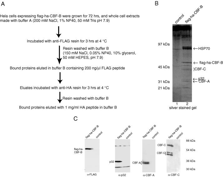
Figure 1.

Purification and characterization of CBF-B complex. (A) Flow diagram for purification of the CBF-B complex from HeLa cells expressing flag-ha-CBF-B. (B) SDS–PAGE analysis of purified CBF-B complex eluted from anti-HA affinity resin. The lanes 1 and 2 were visualized by silver staining. The two different forms of CBF-A and three different forms of CBF-C subunits are probably not stained equally well with a silver stain. (C) Western blot of the purified CBF-B complex eluted from anti-HA resin with anti-FLAG, anti-p32, anti CBF-A and anti-CBF-C antibodies. Extracts from HeLa cells not expressing flag-ha-CBF-B were fractionated in the same way to serve as the control. In case of the blot with mouse monoclonal anti-p32 antibody, the IgG heavy and light chains, coming from the fractionation through anti-FLAG resin (containing mouse monoclonal FLAG antibody) are also detected.
