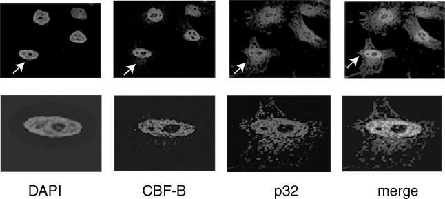
Figure 4.

Colocalization of endogenous CBF and p32 proteins in HeLa cells. This was studied by immunostaining these proteins with their specific primary antibodies and fluorescent-labeled appropriate secondary antibodies. The nuclei were stained with DAPI. The red staining corresponds to p32 protein while the green corresponds to of CBF-B. The merge shows an overlay of red and green fluorescence data. The cell marked by arrow in the upper panel is shown in the lower panel for better details.
