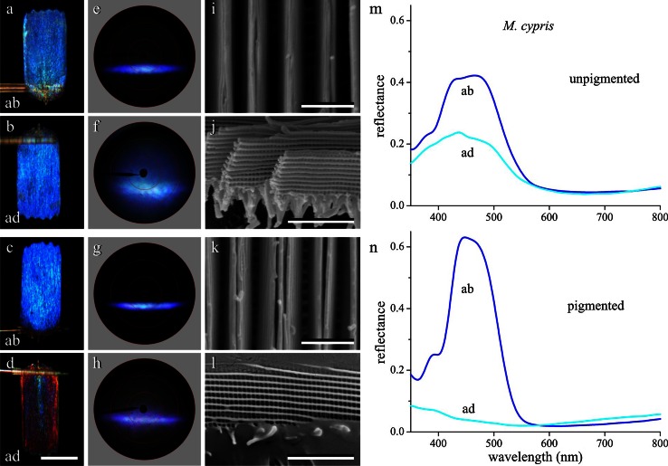
Fig. 4.
An unpigmented and pigmented scale of M. cypris. a, b Micrographs of an unpigmented single scale, abwing (ab) and adwing (ad). c, d Micrographs of a pigmented single scale. e, f Scatterograms of the unpigmented scale. g, h Scatterograms of the pigmented scale. i, k SEM of the upper lamina in normal view of an unpigmented and pigmented scale, respectively. j, l Lateral view of the large stack of scale lamellae. m, n MSP reflectance spectra of the ab- and ad-wing sides of an unpigmented and pigmented scale, respectively. Bars a–d 100 µm; i–l 2 µm

