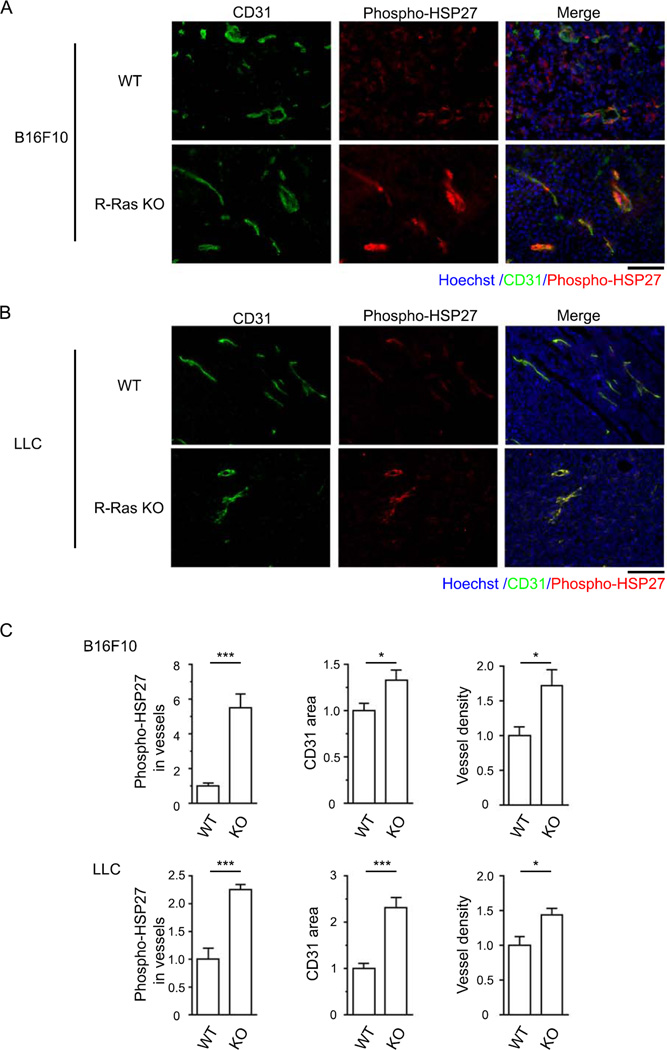
Figure 4. Genetic ablation of R-Ras increases phosphorylation of HSP27 in the tumor endothelium.
B16F10 melanoma (A) or LLC (B) tumors were grown for 10 or 14 days, respectively, in R-Ras KO or wild-type control mice. The tumor endothelium was analyzed for HSP27 phosphorylation by co-immunostaining of tumor sections with anti-CD31 and phospho-HSP27 Ser 78 antibodies followed by microscopy. Scale bar, 100µm. (C) Quantification of HSP27 Ser 78 phosphorylation. The area of phospho-HSP27+CD31+ double staining was quantified and normalized to total CD31 area to analyze phosphorylation of HSP27 in the tumor vessels. The tumor vascularity in each group was determined by total CD31 area and vessel density in tumor sections. Data are presented as fold increase relative to the wild-type control animals. *p<0.05, ***p<0.001. Two animals per group. The total number of vessels analyzed for B16F10 tumors, 258 (WT) and 442 (KO). For LLC tumors, 1239 (WT) and 1774 (KO). Error bars, SEM.

