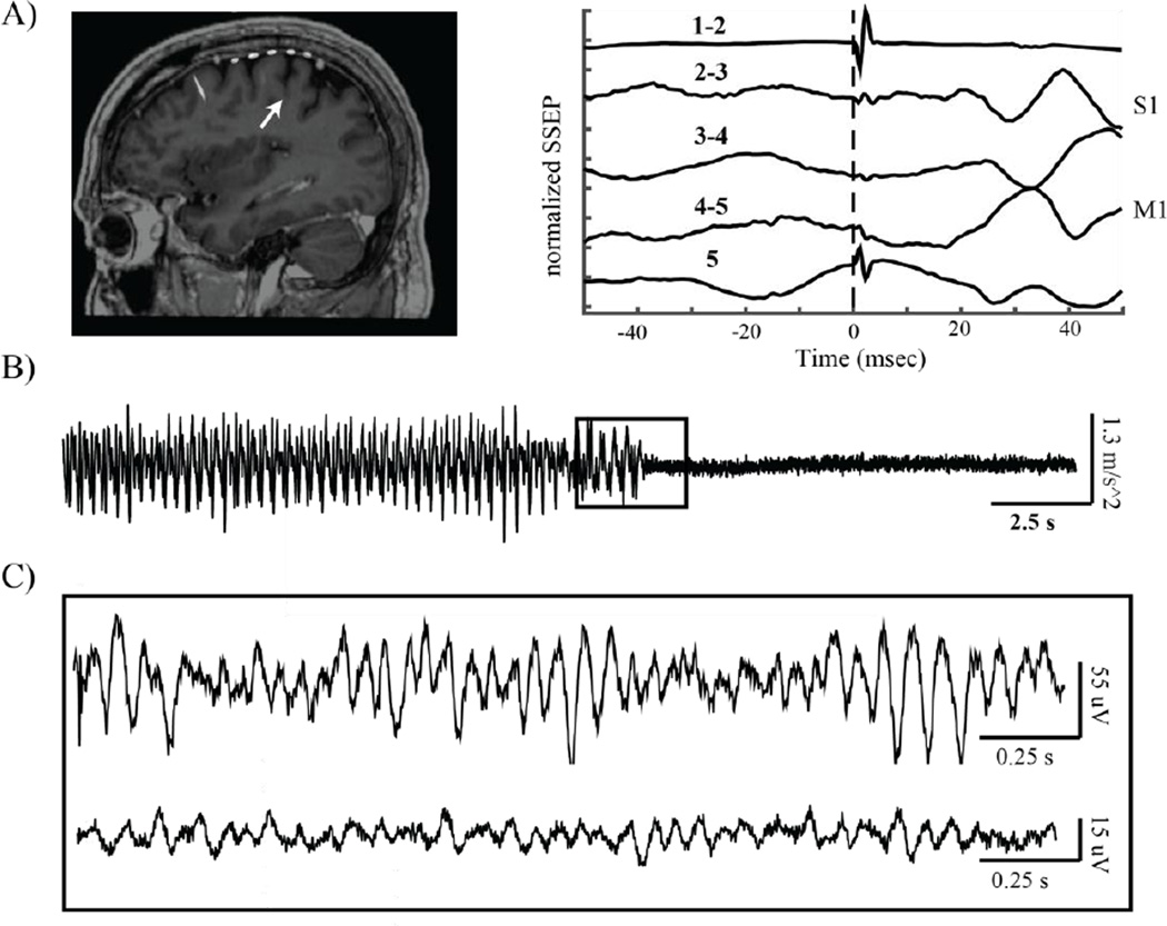
Figure 1.
Data recording methods. A) From left to right: intraoperative MRI/CT fusion showing placement of subdural 6 contact strip over sensorimotor cortex, with arrow pointing to the central sulcus. Median nerve somatosensory evoked potential (SSEP) showing reversal of the N20 potential at the electrode contact pair spanning the primary motor cortex (M1). B) Example accelerometer data obtained from one patient experiencing an epoch of rest tremor, which ended suddenly. C) Example brain recordings corresponding to accelerometer data presented indicated by inset in (B). Top: ECoG data obtained from the contacts spanning M1. Bottom: Simultaneously recorded LFP from two contacts in the dorsal subthalamic nucleus.

