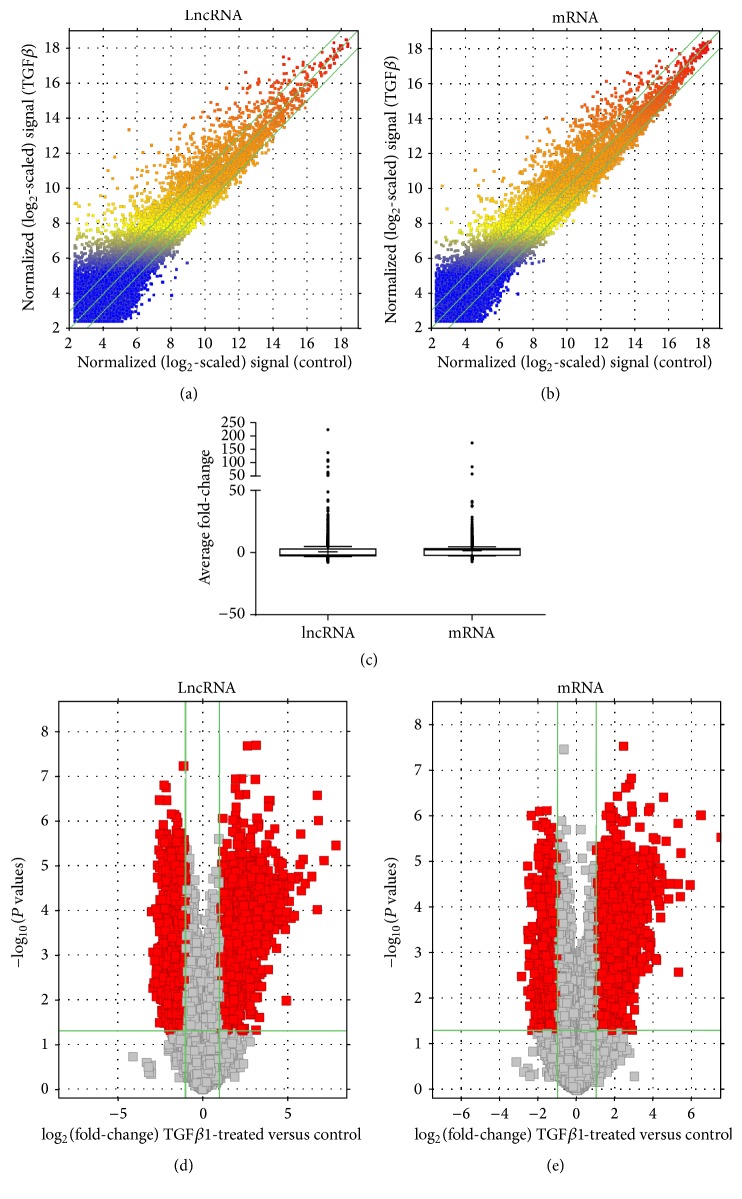
Figure 1.
LncRNA and mRNA expression profiles in HUVECs exposed to TGFβ1 (10 ng/mL) versus control. (a and b) Scatter plots comparing the variation in lncRNA and mRNA expression. The values plotted are the averaged normalized signal values (log 2 scaled) for the control (x-axis) and the TGFβ1 treatment (y-axis) groups. The green lines indicate fold-change. LncRNAs and mRNAs above the top green line and below the bottom green line exhibit at least a 2.0-fold difference between the two study groups. (c) Box-and-Whisker plots (10th and 90th percentiles) showing average fold-change of lncRNAs and mRNAs. Median intensity is denoted with a “−” sign and mean intensity is denoted with a “+” sign. (d and e) Volcano plots detailing magnitude of expression difference. The vertical green lines correspond to 2.0-fold upregulation and 2.0-fold downregulation of expression. The horizontal green line indicates a P value of ≤0.05. Red points represent lncRNAs and mRNAs with statistically significant differential expression (fold-change ≥ 2.0, P ≤ 0.05).

