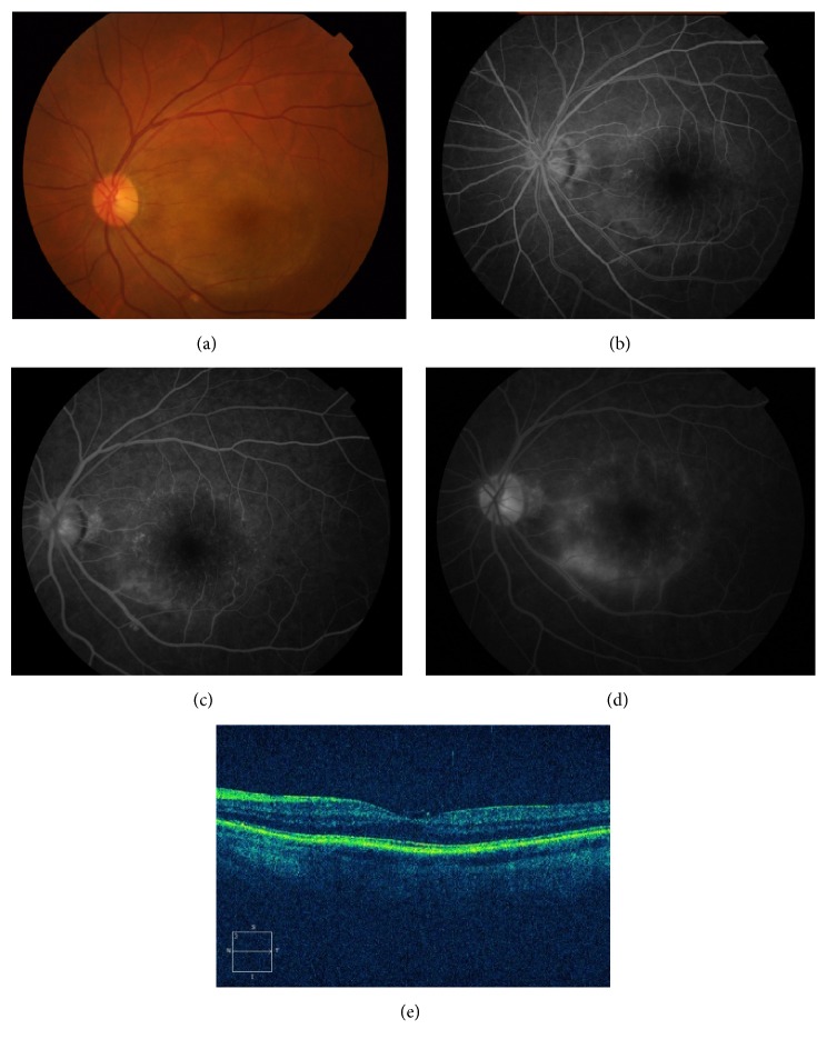
Figure 1.
Patient 1 in the tables with acute syphilitic posterior placoid chorioretinitis. (a) Fundus photograph of the left eye showing a placoid, yellowish, outer retinal lesion. (b) Early-phase fluorescein angiogram showing faint hyperfluorescence in the area corresponding to the lesion. ((c) and (d)) Midphase fluorescein angiogram showing progressive hyperfluorescence followed by late staining. (e) OCT scan illustrating partial ill-defined IS/OS junction, irregular RPE, and a fine epiretinal membrane on the retinal surface.

