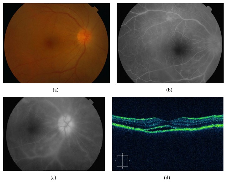
Figure 2.
Patient 11 in the tables with syphilitic retinal vasculitis and papillitis. (a) Fundus photograph of the right eye showing disc and retinal edema. ((b) and (c)) Fluorescein angiogram showing hyperfluorescence with leakage of dye from retinal vessels as well as from optic nerve head. (d) OCT scan illustrating subretinal fluid and irregular RPE.

