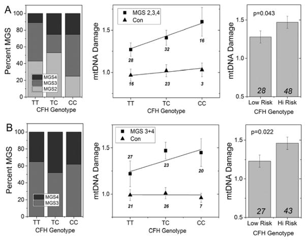
Figure 4. Association Between mtDNA Damage and the CFH Risk Allele. (A).
Refined analysis of data for CFH from Figure 3 shows the distribution of AMD donors (MGS2, 3, 4) for each CFH genotype (left panel) and the mtDNA damage measured in control (n=42) and AMD donors (n=76) (middle panel). A significant linear increase in mtDNA damage correlated with content of the C risk allele in AMD donors (p=0.049) but not in control donors (p=0.59). AMD donors harboring 1 or 2 high risk alleles had significantly higher mtDNA damage compared with AMD donors homozygous for the low risk alleles (TT) (p=0.043; right panel). (B) Distribution of AMD donors (MGS 3 and 4 only) for each genotype (left panel) includes 43 donors from previous studies (Karunadharma et al., 2010; Terluk et al., 2015) and 27 new donors. The extent of mtDNA damage for AMD and control donors for each genotype is shown in the middle panel. While there was a 20% increase in mtDNA damage in donors carrying the C allele, a linear relationship was not observed (p=0.11). Regression analysis of mtDNA damage for control donors (including 12 new donors) was not significant (p=0.09). T-test analysis (right panel) showed AMD donors harboring 1 or 2 high risk alleles had significantly higher mtDNA damage compared with AMD donors homozygous for the low risk alleles (TT) (p=0.022). Total number of donors in each group is indicated on the graph. Data shown are mean ± SEM.

