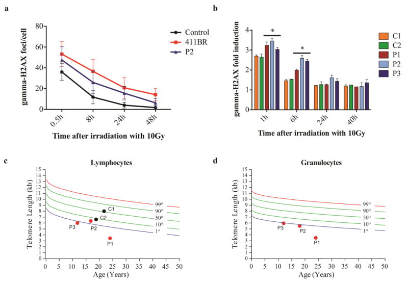
Fig. 2. Increased sensitivity to ionizing radiation (IR) and short telomere length in genotypically affected siblings.
Fibroblasts of P2 were irradiated with 5Gy and γH2AX foci were counted in at least 50 cells using a Fluoview FV 1000 confocal microscope (Olympus) with an UplanApo 60x/1.2na water lens (Olympus) at given time points after IR. Numbers of γH2AX foci/cell are compared to the established LIG4-deficient cell line 411BR [11] and a healthy control (a). PBMCs from patients P1-P3 and two controls C1-C2 (healthy siblings III.2 and III.3) were irradiated with 10Gy, and γH2AX was assessed by flow-cytometry. The γH2AX fold induction was calculated by dividing the mean fluorescence intensities (MFI) of unirradiated cells by the MFI of irradiated cells of each time point (* p≤0.05) (b).
Telomere length in lymphocytes (c) and granulocytes (d) is expressed relative to healthy age-matched controls. The colored lines represent percentile thresholds. In two controls, granulocyte event numbers were too low to measure the telomere length with confidence.

