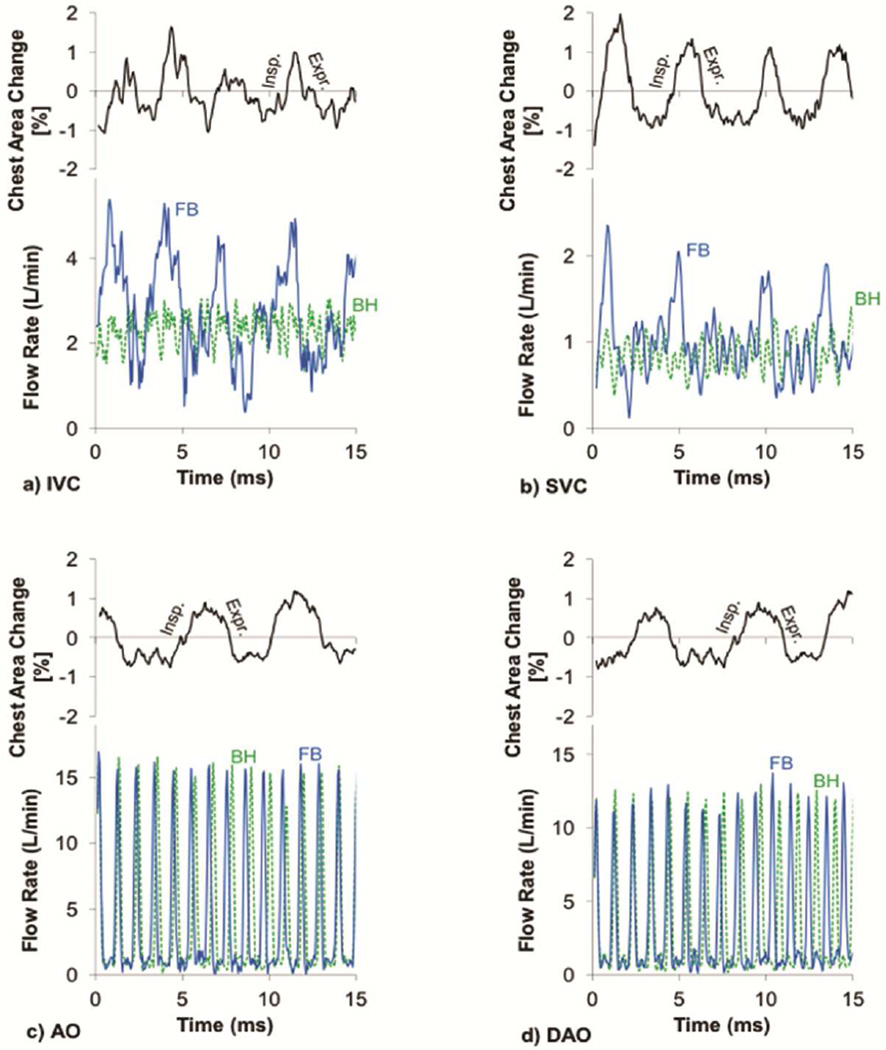
Figure 3.
Resting flow waveforms of a) inferior vena cava (IVC), b) superior vena cava (SVC), c) aorta (Ao), and d) descending aorta (DAo) at the free breathing (solid line) and breath holding (dashed line) conditions along with the corresponding respiratory cycles determined by tracking the chest wall motion for a representative patient

