
Fig. 2.
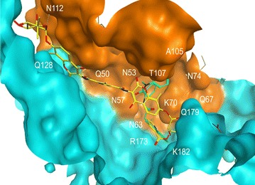
Molecular docking of C-A1 (in yellow) in HIV-1 hexameric capsid structure 4XFZ.pdb with bound PF-74 (in green) using the Compute: SiteFinder option in MOE software. A single C-A1 conformation is shown bound to capsid. The two subunits of capsid are depicted in brown and turquoise respectively. The position of key residues is indicated