Fig. 5.

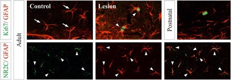
In situ expression of astroglial genes controlled by cAMP in the cerebral cortex in different states. Confocal images of double immunofluorescences of Mki67, Nestin, NR2C and GFAP in the hippocampal dentate gyrus (Mki67/GFAP) and the stratum radiatum of the CA1 region (NR2C/GFAP), and upper layers of neocortex (Mki67/Nestin) of postnatal and adult brains. Cortical lesions were analyzed 5 days after a stab wound injury in the contralateral (control) and ipsilateral (lesion) hemispheres. Arrows point to identified astrocytes expressing only GFAP. Arrowheads identify double labeled cells. Crosses and the asterisk indicate the lateral ventricle and the brain surface, respectively. Scale bar: 10 μm
