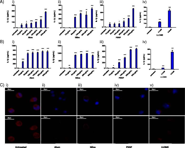
Fig. 1.

Particulates and LLOME induce perturbations in lysosomal compartments leading to the release of IL-1β. ELISA analysis of IL-1β production in supernatants harvested from: a Peritoneal macrophages primed for 3 h with LPS (100 ng/mL) and stimulated for 16 h with a concentration range (10–1000 μg/mL, 22–2200 pg/cell) of i) alum, ii) silica, iii) PSNP, iv) LLOME, b BMDMs primed for 3 h with LPS (100 ng/mL) and stimulated for 16 h with a concentration range (10–1000 μg/mL, 22–2200 pg/cell) of i) alum, ii) silica, iii) PSNP, iv) LLOME. For all experiments n = 4 +/− standard error. c Confocal microscopy of acidified lysosomes labelled with acridine orange (3 μM) in peritoneal macrophages which were i) left untreated or treated for 4 h with ii) 500 μg/mL alum, iii)250 μg/mL silica, iv) 500 μg/mL PSNP, v) 0.5 mM LLOME. Top panel: nuclear dapi stain (blue) + acridine orange (red). Lower panel: acridine orange only. Images representative of 2 independent experiments., scale bar = 20 μm
