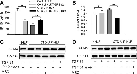
Fig. 7.

Suppression of the myofibroblast phenotype in CTD-UIP HLF through the activation of attenuated TGF-β1 signaling and subsequent IP-10 induction. a, b IP-10 levels (a) and western blot analysis of α-SMA expression (b) in NHLF and CTD-UIP HLF in the absence or presence of TGF-β1. Data are representative of three independent experiments. Representative blots from three replicates are shown. Quantification of α-SMA expression by densitometric analysis was performed using Gel-Pro software. * P < 0.05, ** P < 0.01. c, d Representative western blot for α-SMA expression in CTD-UIP HLF treated with MSC or TGF-β1 in the absence and presence of neutralizing antibody for either human IP-10 (2 ug/ml) (c), or human TGF-β1(1 ug/ml) (d). GAPDH was used as a loading control. Representative blots from three replicates are shown. CTD-UIP-HLF HLF isolated from lung tissues pathologically diagnosed with UIP in CTD-IP patients, HLF human lung fibroblasts, TGF-β1 transforming growth factor-β1, IP-10 interferon γ-induced protein 10, α-SMA α-smooth muscle actin, NHLF normal human lung fibroblasts
