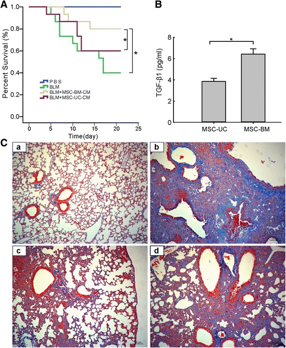
Fig. 8.

Mesenchymal stem cells from bone marrow and umbilical cord exert different efficacy in BLM-induced pulmonary fibrosis mouse model. (A) Survival rates of C57BL/6 mice in the control group and BLM-induced group without any treatment or with treatment by supernatant from either MSCs-BM or MSCs-UC. Supernatants harvested from MSC (1 × 10⁶) culture were intratracheally administered to mice 48 hours after BLM treatment. Analysis was conducted by a logrank test based on the Kaplan–Meier method. (B) An enzyme-linked immunosorbent assay demonstrated a significantly higher level of TGF-β1 secreted from HBMSCs than from MSC-UC. (C) Representative Masson staining photomicrographs of the lung tissue sections from mice 21 days after saline exposure (a), BLM exposure (b), BLM exposure with treatment of the supernatant from MSC-BM (c), and BLM exposure with treatment of the supernatant from MSC-UC (d). 200× magnification. MSCs-BM mesenchymal stem cells isolated from bone marrow, MSCs-UC mesenchymal stem cells isolated from umbilical cord, TGF-β1 transforming growth factor-β1
