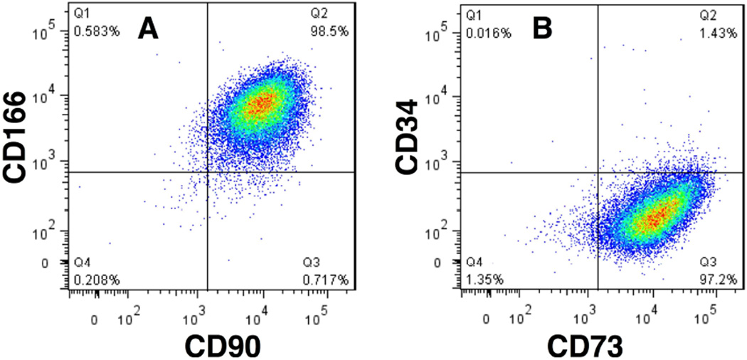
Figure 2.
MSC-surface markers on CSSCs. CSSCs, passage 3, were stained for cell surface antigens and analyzed by flow cytometry. Horizontal and vertical lines show the maximum fluorescence of cells stained with non-specific isotope control antibodies using procedures described by Du et al.17 A. 98% of CSSCs stained for both CD90 (Thy-1) and CD166 (ALCAM). B. 97% of CSSCs stained for CD73 (NT5E), but <2% were positive for the hematopoietic stem cell marker CD34.

