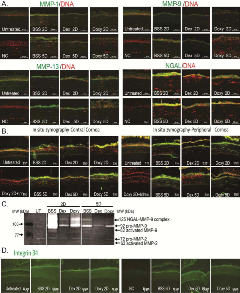
Figure 3.

Anti-inflammatory therapy decreases MMP protein expression and gelatinolytic activity.
A. Representative merged digital images of cornea cryosections immunostained for MMP-1, MMP-9, MMP-13 and NGAL (all in green) with propidium iodide nuclei counterstaining (red) subjected to a combined model of alkali burn and dry eye topically treated with BSS, Dex, or Doxy for 2 or 5 days (2D or 5D, respectively). Scale bar=50 μm.
B. Representative merged digital images of in situ zymography of central and peripheral cornea (green) with propidium iodide nuclei counterstaining (red) in all treatment groups. Doxy 2D was incubated with inhibitor (inh) provided in the kit and used as negative control. Scale bar=50μm.
C. Representative gelatin zymogram showing MMP-9 and NGAL-MMP-9 bands in whole corneal lysates in the treatment groups.
D. Representative digital images of cornea cryosections immunostained for Integrin β4 (in green) in all treatment groups. Nuclear counterstaining was omitted to facilitate visualization of immunofluorescent staining. Scale bar=50 μm.
UT=untreated cornea, BSS=corneas subjected to alkali burn and dry eye and treated topically with balanced salt solution, Dex=corneas subjected to alkali burn and dry eye and treated topically with dexamethasone, Doxy=corneas subjected to alkali burn and dry eye and treated topically with doxycycline, D=days, NGAL=Neutrophil-gelatinase associated lipocalin, NC=negative control.
