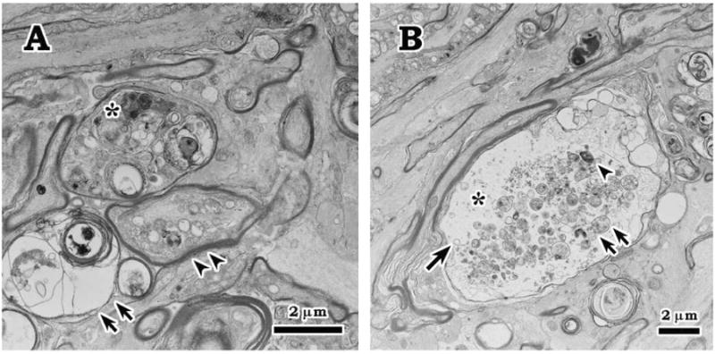
Figure 4.

Axonal degeneration at 7 days after IOP elevation. A. Electron microscopic examination revealed degenerating axons with various features close to the level of lamina cribrosa in the optic nerve head such as accumulation of condensed (*) and swollen (double arrowheads) intra-axonal organelles, and myelin degeneration (double arrows). B. Scattered swollen axon with expanded axoplasm (*) and accumulation of myellin materials (arrowhead), swollen mitochondria (double arrows), and dilated membrane-bounded organelle (arrow).
