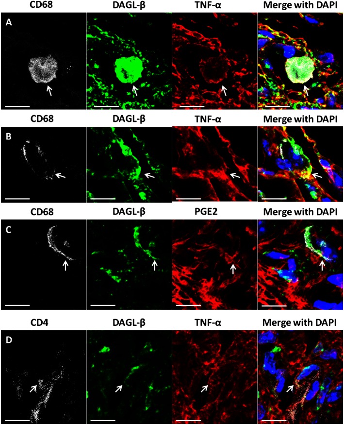
Figure 2.

Qualitative confocal images of cellular immunostaining of DGLβ, TNF‐α and PGE2 in DAGL‐β (+/+) paw pads. (A–D) Paw pad tissue from mice treated with LPS. (A,B) Immunostaining of DGLβ (green) in paw pads is co‐labelled (yellow) with TNF‐α (red) on CD68/ED1 positive (white) cells. DAPI nuclear labelling is blue. Arrows indicate DGLβ and TNF‐α co‐labelling. (C) Immunostaining of DGLβ (green) in paw pads is co‐labelled with PGE2 (red) on CD68/ED1 positive (white) cells, with DAPI nuclear labelling (blue). Arrows indicate co‐labelling of DGLβ and PGE2. (D) Immunostaining of DGLβ (green) in paw pads with TNF‐α (red) on CD4 positive cells, with DAPI nuclear labelling (blue). Arrows indicate co‐labelling of DGLβ and TNF‐α. In all images, the scale bar is equal to 20 μm.
