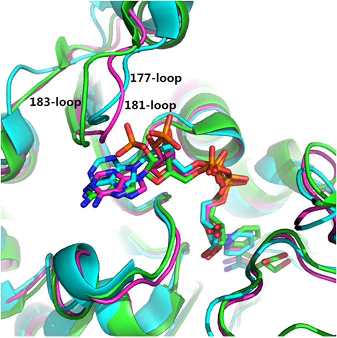
Figure 1. Alignment of three pdb, 1J49 (cyan), 2DBQ (magenta) and 2GCG (green).

The only notable difference between these three binding pocket is at 177-loop of 1J49 (181-loop of 2DBQ, 183-loop of 2GCG). Abbreviations: pdb, protein data ban; 1J49, d-LDH which is NADH-dependent enzyme; 2DBQ and 2GCG, glyoxylate reductases which are NADPH-dependent enzymes.
