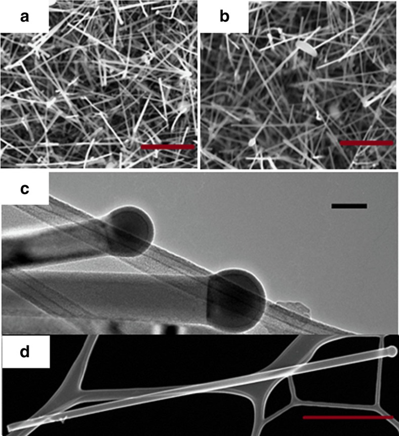
Figure 1. Morphological examination of alloy nanowires by electron microscopy.
SEM images of catalysed Ge1−xSnx nanowires grown using 15 at.% of Sn containing solution with: (a) Au (b) Au0.90Ag0.10 catalysts (scale bar, 1 μm). TEM image in c confirms the participation of VLS growth mechanism with dark-contrast spherical seed at the tip of the nanowire with AuAg catalysts from precursor solution containing 15 at.% Sn. Scale bar, 100 nm. HAADF STEM image in d confirms uniform nanowire diameter along the length with negligible tapering. Scale bar, 1 μm.

