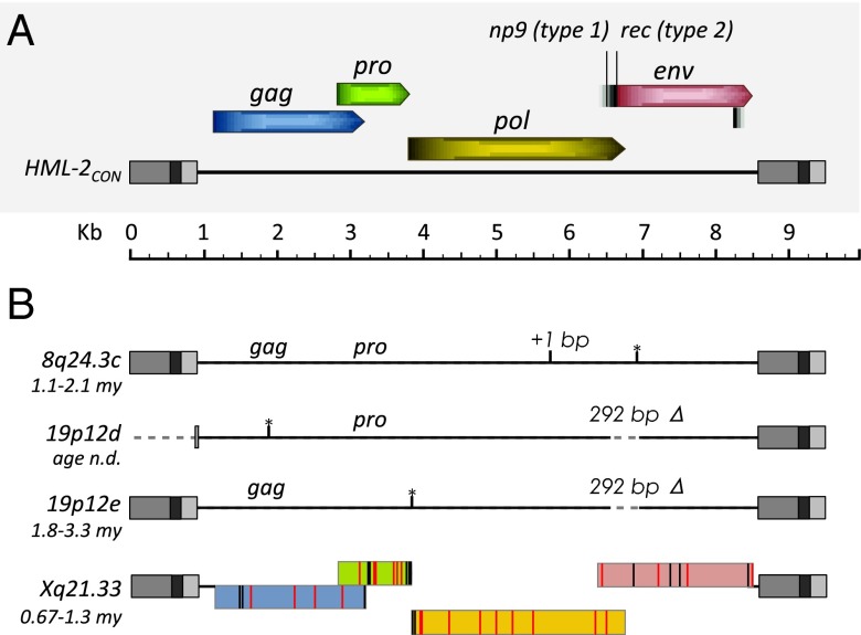
Fig. 4.
Features of newly identified HML-2 proviruses in humans. (A) Schematic representation of the consensus HML-2 provirus, including the viral gene positions and frames to scale. Splice sites for np9 (type 1 insertion, 292 bp Δ) and rec (type 2) are indicated. Regions within the LTRs are colored in gray: U3, medium; R, dark; U5, light. (B) Features of nonreference identified proviruses are shown to scale. The region of 292 bp is labeled for type 1 insertions. Age estimations are shown for each site. n.d., not determined. The black vertical line indicates a frameshift mutation (as indicated “+1 bp”); black lines with asterisks are used to indicate positions of stop codons where present. Reading frames are shown for the Xq21.33 2-LTR provirus as colored as in A. Black vertical lines within the frames indicate the positions of base changes that are observed in other full-length HML-2 proviruses. Red vertical lines are used to indicate base changes that are unique to the sequenced Xq21.33 provirus.

