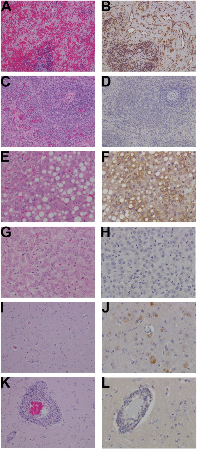
Fig. 4.
Spleen, liver, and brain histopathology of JUNV-infected guinea pigs. A, B, E, F, I, and J are tissues from infected control animal 4–1 (Fig. 3) euthanized on day 14. C, D, G, H, K, and L are from animal 1–2 (treated with J199 on day 6+10) euthanized at study termination on day 40. A, C, E, G, I, and K are tissues stained with hematoxylin and eosin stain (H&E) and B, D, F, H, J, and L are immunohistochemistry (IHC) detecting JUNV antigen. In total, the images demonstrate that the control animal has extensive lesions as visualized with H&E and JUNV-specific antigen is associated with these lesions as determined by IHC. (A) Spleen: diffuse lymphoid depletion, degeneration, and hemorrhage. (B) Spleen: diffuse cytoplasmic immunolabeling of mononuclear cells and endothelium for JUNV antigen (brown). (C) Spleen: no significant lesions. (D) Spleen: no significant immunolabeling. (E) Liver: diffuse hepatocellular vacuolar degeneration and kupffer cell hyperplasia. (F) Liver: punctate cytoplasmic immunolabeling of hepatocytes and kupffer cells. (G) Liver: reactive kupffer cell hyperplasia. (H) Liver: no JUNV antigen detected. (I) Brain: diffuse gliosis. (J) Brain: diffuse cytoplasmic immunolabeling of neurons. (K) Brain: reactive lymphocytic perivascular cuffing and diffuse gliosis. (L) Brain: no JUNV antigen detected. (Magnification: A–D, I, and K, 20x; E–H, J, and L, 40x.)

