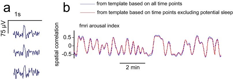
Fig. S8.
Effects of removing potential sleep epochs. (A) Example of a vertex sharp wave identified in one epoch of the LFP data, shown across three electrodes in parietal cortex. (B) fMRI arousal index, as estimated from template maps computed either on all time points (blue) or on the set of time points excluding potential sleep and sleep events (red). Data are shown for a time interval in which no potential sleep or sleep events were identified.

