Fig. 4.
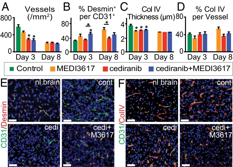
Dual cediranib+MEDI3617 therapy improves vessel normalization in U87 tumors as compared with cediranib monotherapy. U87 tissues were collected from mice treated with control (green bars), MEDI3617 (orange bars), cediranib (red bars), or dual cediranib+MEDI3617 (blue bars) therapy at days 3 or 8 after beginning treatment. Sections were stained for CD31, either desmin or collagen IV (BM), and DAPI. (A and C) Both MVD (A) and BM thickness (C) were significantly decreased at day 3 by both cediranib and dual therapy as compared with control treatment and remained low at day 8. (B) In the dual therapy-treated tumors, perivascular cell coverage (the percentage of the desmin/CD31 double-positive area in the CD31+ area) also was significantly higher on day 3 as compared with control tumors. (D) There was no significant difference in BM coverage among groups. (E) Representative images of CD31 (green)/desmin (red) staining in the normal brain (nl brain) and in control (cont)-, cediranib (cedi)-, and dual therapy (cedi+M3617)-treated tumors. (F) Representative images of CD31 (green)/collagen IV (red) staining in the normal brain and in control-, cediranib-, and dual therapy-treated tumors. Error bars represent the SEM. *P < 0.05 compared with control unless otherwise indicated. (Scale bars, 50 μm.)

