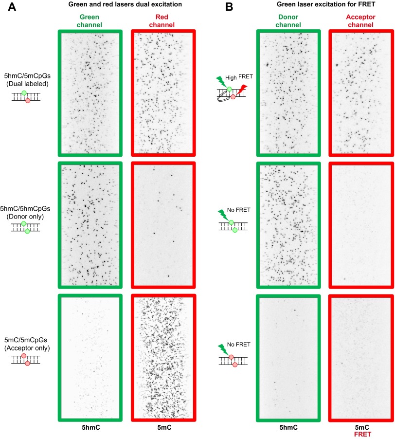
Fig. S6.
Example images of model DNA with different CpG sites. (A) Green and red lasers dual excitation and (B) green laser excitation-only smFRET experiments of model DNA with different CpG sites. (Top) Example image of model DNA containing a 5hmC/5mCpG site. (Middle) Example image of model DNA containing a 5hmC/5hmCpG site. (Bottom) Example image of model DNA containing a 5mC/5mCpG site.

