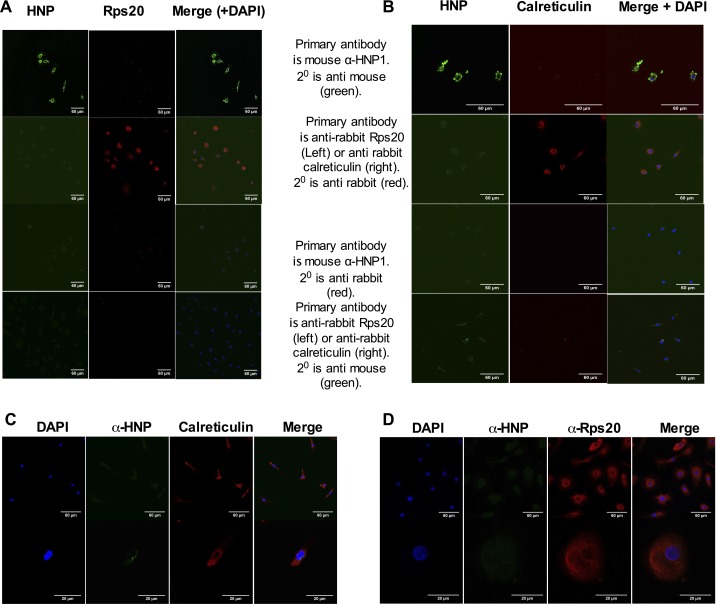
Fig. S3.
(A and B) Confocal microscopy images of HNP1-treated HMDMs. As staining controls, anti-HNP1–3, anti-Rps20, or anti-calreticulin primary antibodies were followed by the “wrong” secondary antibodies (either anti-rabbit or anti-mouse secondary antibodies, respectively). DAPI (blue) is seen on the merged images. HMDMs that had not been treated with HNP1 were stained with primary and secondary antibodies to HNP1–3, (C) Rps20, or (D) calreticulin as indicated. Nonspecific staining of HNP1 is not seen. n = 3.

