Fig. 3.
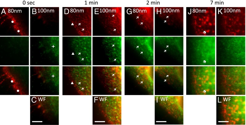
Multiangle-TIRF imaging of EGF-activated clathrin-mediated endocytosis. (A–C) A431 cells were incubated with Alexa Fluor 555-labeled EGF ligand (red) for 1 h at 4 °C. (D–L) Cells were shifted to 37 °C for 1 min (D–F), 2 min (G–I), and 7 min (J–L). Cells were fixed and stained with antibodies to clathrin and with secondary antibodies conjugated to Alexa Fluor 488 (green). Cells in A–C were fixed immediately after the 4 °C incubation with fluorescent EGF (0 s). Dots in A mark the EGFR-ligand complex located at the plasma membrane, seen at the 80-nm layer at time 0 s. The EGFR-ligand complex was found in clathrin-coated pits and vesicles at the plasma membrane and inside the cell at 100 nm and beneath after the 1- to 2-min incubation at 37 °C (D, E, G, and H, arrows). The EGFR-ligand complex was found in endosomes after 7 min (double arrows in J). Images were collected using the image-photobleach-image protocol at penetration depths of 80–100 nm. Wide-field (WF) images (C, F, I, and L) were collected as well. (Scale bars: 5 μm.)

