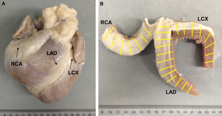
Figure 1.

Preparation of coronary artery sections. A, Heart from an LDLR −/− pig after formalin fixation. B, Coronary arteries with surrounding tissues removed from the heart; yellow lines indicate the 1‐cm intervals at which the coronary arteries were sectioned. LAD indicates left anterior descending coronary artery; LCX, left circumflex coronary artery; RCA, right coronary artery.
