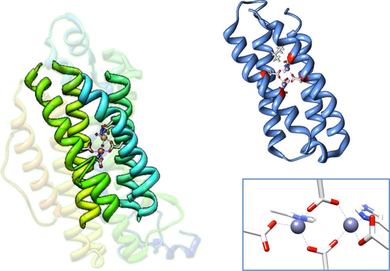
Figure 1.
Crystal structure of ribonucleotide reductase (left) (PDB 1RIB)43 with the four-helix bundle motif bolded and the NMR structure of Zn-substituted DFsc (right) (PDB 2HZ8).44 A closeup of the active site with 2 histidines + 4 carboxylate coordination is shown in the bottom right. The 4A’s shown in red of the NMR structure of DFsc are mutated to 4G’s (A10G, A14G, A43G, A47G) in G4DFsc.

