
Fig. 1.
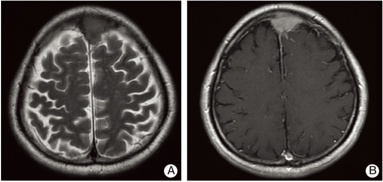
Magnetic resonance imaging of the brain. (A) T2 weighted image shows the 1.9×3.6 extra-axial mass with broad based dural attachment to anterosuperior aspect of falx cerebri involving superior sagittal sinus. (B) T1 weighted image. The mass showed dural tail sign and a suspicious cerebrospinal fluid cleft sign.