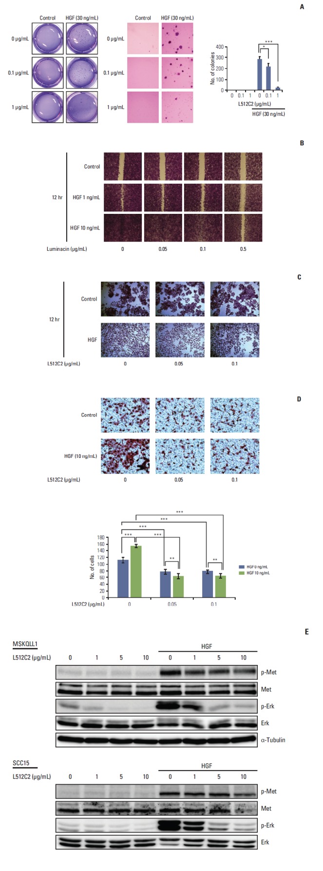
Fig. 3.

Effect of luminacin on hepatocyte growth factor (HGF)–induced viability, migration, and invasion capability in head and neck carcinoma cells. Investigation of cell viability, migration, and invasion capability after HGF/luminacin treatment using colony forming, wound healing, cell scattering, and invasion assays. (A) HNE1 cells were treated or untreated with 30 ng/mL HGF and luminacin (0, 0.1, or 1 μg/mL) and incubated for 3 days to form colonies. After 4% crystal violet staining, more than 2 mm of the colonies were counted. Luminacin treatment significantly inhibited the survival rates of cells treated with HGF. (B) Confluent monolayers of MSKQLL1 cells were wounded by scratching the surface as uniformly as possible with a 1-mL pipette tip. Cells were treated or untreated with 10 ng/mL HGF and luminacin (0, 0.05, 0.1, or 0.5 μg/mL), and then cultivated for another 12 hours. Percent closure of wound areas was measured. Luminacin treatment resulted in significant dose-dependent inhibition of HGF-induced enhancement of cell proliferation and migration. (C) MSKQLL1 cells were cultured with or without luminacin (0, 0.05, or 0.1 μg/mL) and HGF (10 ng/mL). After 24 hours, the cells were stained in a 0.1% crystal violet solution. The number of cell colonies, their sizes, and the degree of scattering were observed. HGF/luminacin co-treated cells showed a discernible reduction in HGF-induced cell scattering. (D) MSKQLL1 cells (2×10⁴) in the upper chamber were pretreated with luminacin (0, 0.05, or 0.1 μg/mL) and then with or without 10 ng/mL HGF. After 16 hours, the number of invading cells was counted in four representative fields per membrane. Co-treatment with luminacin and HGF significantly inhibited cell invasion compared to treatment with HGF alone. (E) Immunoblot of luminacin/HGF treated cells stained with antibodies against Met, p-Met, Erk, and p-Erk. Luminacin inhibited HGF-induced enhancement of phosphorylation of p-Met and the downstream target, p-Erk. Data represent the mean±standard deviation of three independent experiments. *p < 0.05, **p < 0.01, ***p < 0.001.
