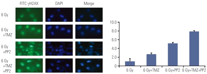
Fig. 2.
Representative images of γH2AX foci formation in U251 cells. γH2AX foci formation was measured in U251 cells after treating PP2 (10 μM), temozolomide (TMZ, 25 μM), or both. The nuclei stained by DAPI are shown in blue, while the γH2AX foci stained by FITC are shown in green. Increased accumulation of γH2AX foci after treating PP2 and TMZ was confirmed by indirect immunofluorescence 6 hours after radiotherapy (6 Gy), indicating delayed DNA double strand breakage repair. All differences between the groups (6 Gy vs. 6 Gy+TMZ, p=0.014; 6 Gy+TMZ vs. 6 Gy+PP2, p=0.020; 6 Gy+PP2 vs. 6 Gy+TMZ+PP2, p=0.016) were statistically significant.

