Fig. 1.

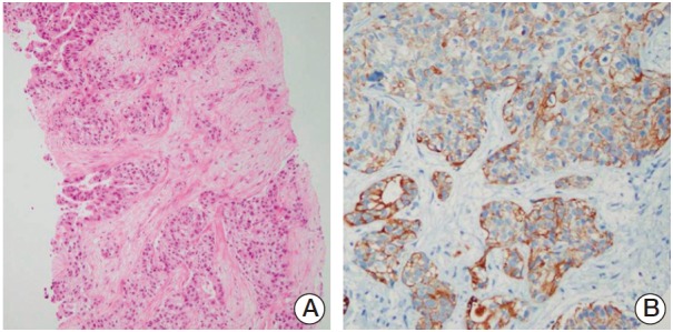
Photomicrographs of the liver biopsy. (A) The liver biopsy indicated adenocarcinoma (H&E staining, ×100). (B) The tumor cells were positive for cytokeratin 19 (×200).

Photomicrographs of the liver biopsy. (A) The liver biopsy indicated adenocarcinoma (H&E staining, ×100). (B) The tumor cells were positive for cytokeratin 19 (×200).