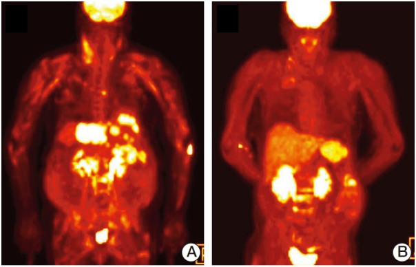
Fig. 2.

Representative [18F]-fluorodeoxyglucose (FDG) positron emission tomography–computed tomography (PET-CT) images of the patient. Whole-body PET-CT was performed using FDG, scanning started 60 to 90 minutes after tracer injection, and images obtained in transverse, coronal, and sagittal planes were reconstructed. (A) Baseline maximum intensity projection (MIP) image. (B) MIP image after two cycles of chemotherapy.
