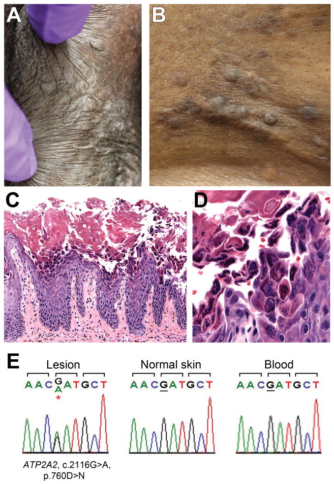
Figure 1.
Clinical, histopathological and sequencing findings.
Top panel: Skin-colored papules on the left external labia majora (a) and central chest (b). Central panel: Hematoxylin and eosin stains of lesional tissue at low power (c) and high power (d) show acanthosis and papillomatosis, with acantholysis and dyskeratosis of keratinocytes. Lower panel: Chromatograms (e) demonstrate mutation of ATP2A2 in affected epidermis with wild-type sequence in unaffected epidermis and peripheral blood.

