
Fig. 2. Hepatocyte-derived exosomes induce hepatocyte proliferation in vitro.
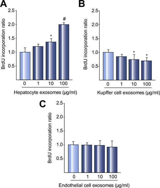
Exosomes isolated from primary hepatocytes (A), Kupffer cells (B), or sinusoidal endothelial cells (C), were added to primary hepatocytes and proliferation measured 24 h later by BrdU incorporation. Exosomes from primary hepatocytes dose-dependently increased hepatocyte proliferation. Kupffer cell exosomes reduced hepatocyte proliferation and exosomes from sinusoidal endothelial cells had no effect on proliferation. For all panels, data are mean ± SD with n = 8 (A, B) or n = 6–8 (C) per group. *p <0.05 compared to media control (0 exosomes), #p <0.05 compared to all other groups.