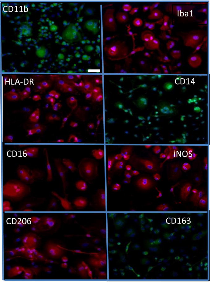
Figure 5.

Cells in DUOC-01 product express a variety of macrophage/microglial antigens. All images are prepared for immunocytochemistry with antibody to the antigen indicated and counterstained with DAPI. Immunostaining is red or green; DAPI, blue. All images are at same magnification. White scale bar in CD11b image is 50 μm.
