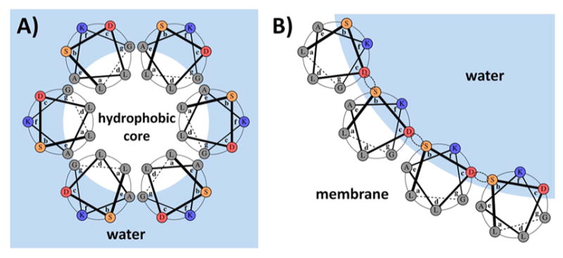
Figure 3.

Design concept illustrated using one of the designed sequences (PSPF-DKG). Hydrophobic residues are either lining the core of the bundle in the water-soluble state (A) or are facing the lipid membrane in the membrane channel state (B). Dotted circles illustrate potential hydrogen bonding in the channel state. Heptad positions in both panels are labeled according to the water-soluble state. The amino acid choices at each position are shown in Table 1.
