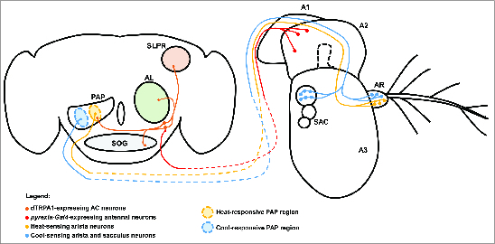
Figure 4.

Central and peripheral neural mechanisms for detecting changes in temperature. The adult Drosophila brain and antenna are schematized here, with colored circles representing neuronal cell bodies, colored lines representing axonal connections, and colored diamonds representing presynaptiC-terminals. Heat-responsive and cool-responsive regions of the proximal antennal protocerebrum (PAP) are schematized as filled ovals with dashed outlines. Heat-sensing neurons of the aristae (AR) connected to the third antennal segment (A3) send projections to the brain that synapse in the heat-sensitive region of the PAP. Cool-sensing neurons of the aristae and sacculus (SAC) send projections to the cool-responsive region of the PAP. Putative pyrexia-Gal4-expressing neurons in the second antennal segment (A2) project to the brain and synapse on the dTRPA1-expressing AC neurons (it is important to note that the exact location and identity of these neurons are unknown). The AC neurons project to the heat-responsive region of the PAP, as well as to the suboesophageal ganglion (SOG), the superior lateral protocerebrum (SLPR), and the antennal lobe (AL). Also depicted is the first antennal segment (A1).
