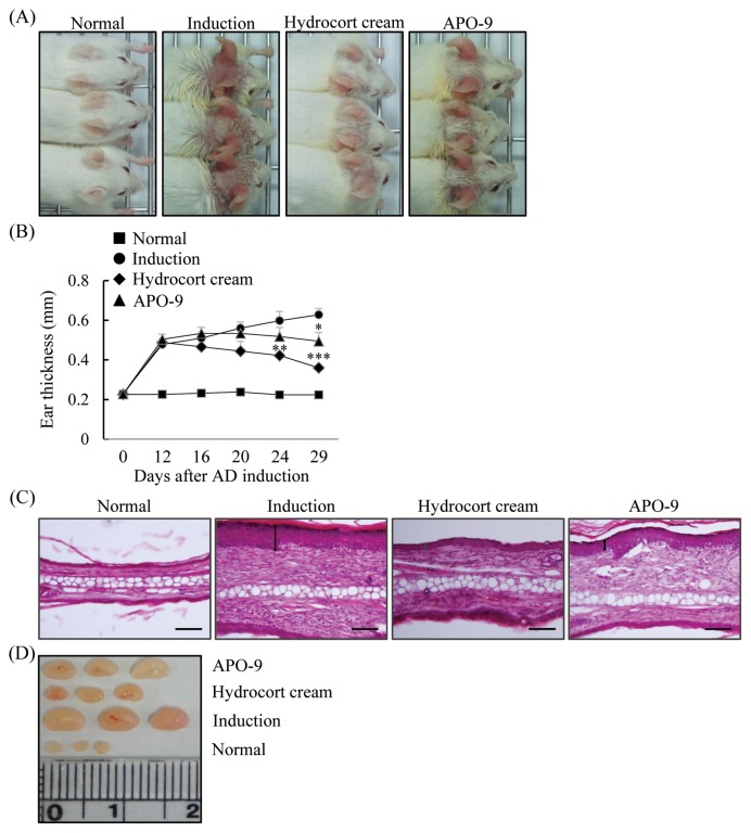
Fig. 4.
Apo-9′-fucoxanthinone suppresses experimental atopic dermatitis. (A) Macroscopic views of the ears and (B) ear thickness measured on days 0, 12, 16, 20, 24, and 29. (C) Paraffin-embeded sections of ear tissue stained with hematoxylin and eosin. (D) The lymph nodes (LNs) were photographed to record morphologic changes (n = 5 mice per group). Scale bar = 0.1 mm. Values represent the mean ± SD. *P< 0.05; **P<0.01 and ***P< 0.001 compared to mice stimulated with DNCB alone (induction group).

