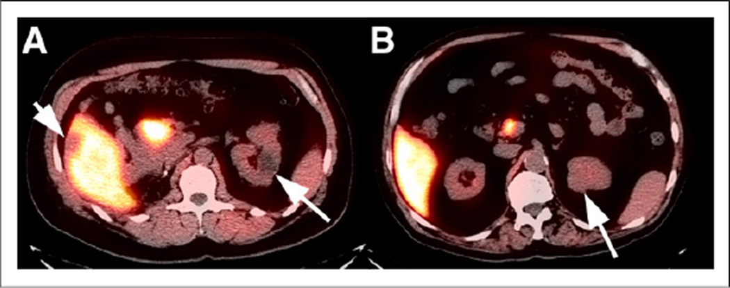
FIGURE 4.
(A) Coregistered axial 18F-FACBC PET/CT image from summed dynamic 18F-FACBC renal study demonstrates lack of uptake in MR imaging–proven hepatic hemangioma (small arrow) and left renal cyst (large arrow). (B) Coregistered axial 18F-FACBC PET/CT image demonstrates uptake in renal cancer (arrow) similar to that of renal parenchyma on summed dynamic images.

