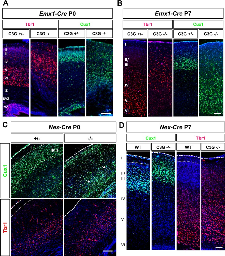
Fig 2. The loss of C3G leads to defects in cortical lamination.
(A-D) Coronal sections from the brains of P0 (A, C) or P7 (B, D) C3GEmx1-KO or C3GNex-KO embryos with the indicated genotypes were stained with antibodies for Tbr1 (layer V/VI and SP; red) and Cux1 (layers II/III, green). Both markers revealed a severe disruption of cortical organization in the C3GEmx1-KO. (B) Staining for Tbr1 and Cux1 at P7 demonstrates that the cortical plate is inverted in the C3GEmx1-KO. (C, D) No severe defects were observed in C3GNex-KO, except for the presence of ectopic Cux1+ cells in deep layers at P0 (marked by arrows) and the invasion of Cux1+ cells into layer I. Dorsal is to the top. Single confocal planes are shown. Scale bars are 100 μm. Images are representative for 3 independent experiments with 3 embryos per genotype from different litters.

