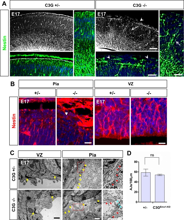
Fig 3. C3GEmx1-KO shows defects in RGCs at the pial surface.
(A, B) Coronal sections of C3GEmx1-KO heterozygous or homozygous knockout embryos were stained with an anti-nestin antibody (green (A) or red (B)) and Hoechst 33342 (blue) at E17. (A) Higher magnification images from the pial surface show a continuous arrangement of glial fibers and endfeet in the heterozygous control compared to the disrupted organization of glial fibers and a rupture of basement membrane in the C3GEmx1-KO cortex (arrowheads). Arrows indicate the disorganized glial fiber network. No defects at the VZ were seen in C3GEmx1-KO (B). Single confocal planes are shown. (C, D) Coronal sections from the C3GEmx1-KO cortex were analyzed by electron microscopy at E17. Images of the pial surface show a distinct BM in heterozygous embryos (yellow arrowheads) with a compact arrangement of cells below the BM (C). The BM appears to be ruptured in the C3GEmx1-KO cortex and the cells protrude outside. The area marked by a box is shown at a higher magnification on the right depicting the BM. The red arrows (magnified panels) mark the continuous BM. The broken BM in the C3GEmx1-KO pial surface is marked by blue arrowheads. No defects were seen at the VZ and the AJs formed normally (C). (D) The number of AJs per 100 μm of the VZ was quantified and no significant differences were found between the heterozygous and homozygous C3GEmx1-KO embryos (n = 3 embryos per genotype, means ± s.e.m., ns, not significant, Student’s t-test). Scale bars are 100μm (A) 50μm (magnified panels in A), 10 μm (B), 500 nm (C, VZ) and 2 μm (C and magnified panels, Pia). Images are representative for 3 independent experiments with 3 embryos per genotype from different litters.

