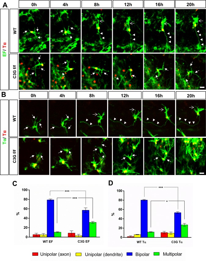
Fig 7. C3G is required in multipolar neurons for neuronal polarization.
(A-B) Wild type (WT) or Rapgef1flox/flox (C3G f/f) E13.5 brains were transfected by ex vivo electroporation with (A) pEF-Cre, pEF-LPL-LynN-EGFP and pTα-LPL-H2B-RFP or (B) pTα-Cre, pTα-LPL-LynN-EGFP and pTα-LPL-H2B-RFP to specifically inactivate the conditional alleles and label early post-mitotic neurons. Imaging was performed 30h after electroporation. Neurons from WT coronal slices first extend a long trailing process followed by a leading process. Slices from Rapgef1flox/flox brains showed a significant number of neurons that remained multipolar and did not extend a trailing or a leading process after more than 20 h of imaging. (C, D) The percentage of cells that formed only a trailing axon (unipolar (only axon), red), only a leading process (unipolar (only leading process), yellow), that became bipolar (blue), or remained multipolar (green) after transfection of pEF-Cre (C) or pTα-Cre (D) at the end of the imaging period of 20 h (means ± SEM, ***p ≤ 0.001, *p ≤ 0.05 two-way ANOVA with Tukey’s multiple comparison test; number of bipolar or multipolar neurons from Rapgef1flox/flox slices compared to control slices; n = 37 (wildtype; EF), n = 29 (Rapgef1flox/flox; EF) and n = 51 (wildtype; Tα), n = 45 (Rapgef1flox/flox; Tα) from 3 independent experiments that each included multiple slices from different animals, n indicates the total number of neurons analyzed in all experiments). The VZ is to the bottom and the pial surface to the top. Scale bars are 20 μm.

